a positron emission tomography pet scan depicts brain activity by detecting:
 Brain positron emission tomography - Wikipedia
Warning: The NCBI website requires JavaScript to operate. Evaluation of brain activity related to the task: Is there a role for the 18F FDG-PET image? Agostino Chiaravalloti 1Department of Biomedicine and Prevention, Faculty of Medicine and Surgery, University of Tor Vergata, Rome, Italy 2IRCCS Neuromed, UOC Nuclear Medicine, Pozzilli (IS), ItalyAlessandro Micarelli 3Department of Clinical Sciences and Translator Medicine, Faculty of Medicine and Surgery, Tor Vergata University, Rome, Italy Maria Ricci 4 Department of Radiological, Oncology and Pathological Sciences, Faculty of Medicine and Surgery, University of La Sapienza, Rome, Italy 5Institute of Cognitive Sciences and Technologies, CNR, Rome, ItalyGabriele Ciccariello 2IRCCS Neuromed, UOC Nuclear Medicine, Pozzilli (IS), ItalyErnesto Bruno 3Department of Clinical Sciences and Translator Medicine, Faculty of Medicine and Surgery, Tor Vergata University, Rome, Italy Marco Alessandrini 3Department of Clinical Sciences and Translator Medicine, Faculty of Medicine and Surgery, Tor Vergata University, Rome, ItalyOrazio Schillaci 1Department of Biomedicine and Prevention, Faculty of Medicine and Surgery, University of Tor Vergata, Rome, Italy 2IRCCS Neuromed, UOC Medicina Nucleare, Pozzilli (IS), ItalyAbstractPositron emission tomography (PET) with 2-[18F]-fluorodeoxyglucose (FDG) has been widely used for the evaluation of cortical glucose metabolism in several neurodegenerative disorders while its potential role in the evaluation of cortical and subcortical activity during a remains brain path. Few studies have been carried out to investigate the potential role of this radio-treater to evaluate the use of brain glucose during the dynamic activation of the brain. The aim of this test is to provide an overview of FDG-PET applications in the evaluation of cortical activation at rest and during tasks, first describing the physiological basis of FDG distribution in brain and its kinetic in vivo. An overview of image and image interpretation protocols will also be offered. As a last aspect, the results of the major studies in this field will be summarized and the results of PET findings made in healthy subjects and patients with various diseases will be reported.1. IntroductionGlucose is the main source of energy for the brain but, to date, the dynamic response of glucose use to changes in brain activity is not yet fully understood. The brain represents only 2% of the body weight, but receives 15% of the heart output, 20% of total body oxygen consumption and 25% of total body glucose use []. For a long time it was assumed that changes in the flow of brain blood (CBF) and in the metabolic rate of oxygen (CMRO2) were closely coupled in both active and resting brain states []. To date, the "neurovascular contamination" (where vasoactive metabolic products such as lactate, CO2/H+ or adenosine are responsible for increased blood flow after increased glucose consumption) has recently been replaced by the "neuronal" hypothesis. The demand for neuronal energy is communicated to the vasculatura in advance, in the form of food by vasoactive neurotransmitters or synaptic signaling products. Therefore, vasodilation occurs independently of the signaling induced by the metabolism of glucose []. The brain needs an abundant and constant supply of oxygen. At rest, 80-92% adjusts to the adenosine triphosphate hails of the oxidative metabolism of glucose; therefore, a functional activation, which implies the need for additional ATP and oxygen, must cause a couple increase both CBF and CMRO2 [, ]. However, previous studies on oxidative metabolism, performed with magnetic resonance (MRI) or postitron emission tomography (PET) with 15O radiotrators tagged, reported that large increases induced by stimulus in the FBC were accompanied by a small increase of CMRO2. These data indicate that, during the short-term functional activation, CBF and CMRO2 are not directly linked [, ]. Postitron emission tomography (PET) with [18F]-fluorodeoxyglucosa (FDG) allows the quantitative measurement of the cerebral metabolic rates of glucose (CMRGlu). Due to its high spatial and temporal resolution, its extensive distribution and radioprotection advantages compared to the PET image, the MRI remains in our opinion the mode of choice of image for functional activation studies. However, the aim of this review is to explore the current knowledge of the PET-FDG image in task activation studies due to the ability of PET to explore in different biochemical aspects the possibility of doing this test in subjects with contraindication to the MRI. Brain tissue can be considered as a three-component model. The deoxyglucose-6-phosphate (DG-6-P), once formed, is essentially trapped in the tissue during the duration of the experimental procedure, allowing to obtain images of the FDG kinematic in the brain 30 minutes after the injection [–]. In particular, taking into account the "meseta" tendency of the 18F-FDG kinetics, 40% of the radial compound is extracted into the brain (~250 nCi/g) in the first minutes after the injection allowing the detection of the areas of the cortical brain that are activated in the early times of the task [–]. Therefore, FDG-PET has been considered a useful diagnostic tool, especially in the population that presented contraindication to MRI (such as subjects with metallic devices (clips, vascular stents, heart devices and cochlear implants), foreign bodies, tattoos containing metal particles, or claustrophobia []). Despite previous documents on the physiological magnitude response to stimuli during hypoglycemia suggest otherwise [], most authors assumed that the use of glucose can change in parallel with the FBC and during neuronal activation. However, it is well known that in humans physiological stimulation results in a dramatic increase in brain blood flow and CMRGlu, especially in short-term stimulation []. Instead of this dramatic increase of CMR Glu in short-term stimulation, previous works focused on the effects of continuous visual stimulation described as well as the rapid increase of CMRGlu. This increase of CMRGlu was followed by attenuation of this trend after 20 minutes of continuous stimulation that can reflect a change of glucolytic glucolysis to oxidative glucose metabolism with continuous activation []. To quantify the capture and metabolism of the tracer in tissue, a cynical model of FDG in PET image, whose efficiency is demonstrated in physiological conditions in the brain, is crucial for a correct methodological approach to PET activation studies. DG-6-P, once formed, is trapped in the tissue for a reasonable time allowing to obtain images of the brain after injection and therefore FDG-PET can be considered as a crucial tool in evaluating brain activity related to tasks [–].2. Exploration Procedure The measurement of CMRGlu represents neuronal processes after an intravenous injection of FDG []. Most PET image activation studies aimed at evaluating CMRGlu changes during the tasks required a separate photo shoot for rest and task activation conditions. The execution of two scans leads to additional radiation with important implications in terms of radioprotection. Therefore, a new approach that required a single acquisition of scan seems very convenient. The CMRGlu, according to this procedure, can be measured on the basis and after stimuli during the same session, improving the evaluation of the specific metabolism of the task. Taking into account the intrasubjective variability between repeated scanning, caused by several events (e.g. caffeine, sleep condition, changes in blood chemistry and inregistration), a single scanning procedure can increase both the sensitivity and the specificity of the activation Image PET []. Obviously, this approach presents new undeniable advantages in terms of costs, radioprotection and patient compliance (assuming that the specific task can be done in the nuclear medicine unit). The widely used clinical use method, used both in individual studies and in two PET imaging scanners, is characterized by a scan made 20-40 minutes after a PDG bolt injection. The Bolus method can provide a quantitative and/or qualitative measurement of the CMRGlu after stimuli. In fact, in most of the previous activation, PET studies used an FDG bolus injection to characterize functional metabolic responses to multiple and different types of stimulation, including visual, hearing, cognitive and drug administration tasks. This bolt method reports an integral part of neuronal processes for 20-40 minutes. However, taking into account the "meseta" trend of FDG's kinetic and that 40% of the radio-labeled compound is extracted into the brain (~250 nCi/g) in the first minutes after injection, the CMRGlu after screw injection can be a mirror, in particular the initial metabolic activation [, ]. Therefore, some limitations on the lack of information on dynamic metabolic changes during scanning and/or during the task tend to affect this method that allows the detection of the areas of the cortical brain that are activated in the early times of our task. Recently, a new technique was proposed with a constant infusion of the MDGs for the entire scan [, ]. The advantages of constant infusion of radioligand are to avoid the prominent absorption of the MDGs in the initial period and to provide radioligand available throughout the implementation of the task. These kinetic aspects can improve the quantification of dynamic metabolic changes in the brain during the task, promoting a unique scanning procedure that allows the measurement at the same session CMRGlu at the base and during the execution of tasks [, ].To minimize inter-individual differences, a statistical analysis of the parenting of parameters (SPM) can be generally useful in the dynamic image of PET considering physiological variations of CM In fact, the authors compared the metabolism of rest glucose to the activation of verbal memory tasks in healthy subjects and described both the metabolic decrease related to age and the sexual differences within the front regions that were most markedly found in the front and cingulated medium areas. This pattern is consistent with some patterns related to the age of affective and cognitive change []. In the activation PET imaging studies you should prefer a quantitative analysis on a qualitative visual analysis of images. An accurate quantification of PET measurements can be obtained by measuring trace concentration by blood sampling. This procedure may result in awkwardness for the patient and may therefore be partially avoided by comparison of the MDG absorption in case studies to that of a group of subjects serving as a control group.3. Procedure for the task To date, FDG-PET image was used for the evaluation of CMRGlu during several and different types of tasks, in order to evaluate a physiological and/or pathological response to stimuli. In . Therefore, the PET image can improve the diagnostic and therapeutic approach and/or evaluate the efficiency of the treatment. Potentially activation PET imageg with FDG may be useful in both fisilogical studies of the brain and in most of the diseases, affecting glucose metabolism and CBF but the procedure should be conformed to specific experiment. The experimental procedure may vary according to the registered population (i.e., healthy subjects, patients affected by various types of brain damage, or affected by neurodegenerative diseases) and types of tasks (visual, hearing, somatic, etc.). Table 1Summary of the most relevant studies on PET image with FDG in different types of tasks. Number of themes Image modality type PETOutcomeReferenceVisual manufacturing5 themes Visual stimulation began 5 minutes before FDG injection and continued throughout the study. Hypermetabolism in the primary and associative visual cortex increased the complexity of the visual scenes. [] Motor task theme1 The volunteer suffered 20 min of motor rest and activation (motorized work, which implied repetitively grasping and dropping the right hand, performed during the first 5 min of the activation period)Hypermetabolism in the primary counterlateral motor cortex, the supplementary motor area and the ipsilateral cerebello (right). Engine strength and position 2 subjects FDG injection has been performed during a force and position task (separated for 7 days, with the elbow flexor muscles in 15% maximum voluntary contraction force)The metabolism of the vessel in the occipital and temporal cortices of the brain during the position task compared to the task of force. [] Volitional Traction Tasks8 Male SubjectsPET-FDG at rest and while swallowing (20 seconds intervals for 30 minutes) in the erect seat positionDuring the ingestion of hypermetabolism in left sensory bark, ceebello, talamo, precuneus, previous insula, left and right side glutin, and left and rear right corricle cortex; metabolism decreased in the premotor cortex Visuospacial Complex/motor task8 male subjects The subjects were subjected to PET-FDG performing a complex visuospacial/motor task (the Tetris computer game), before and after 4-8 weeks of daily practice in TetrisAfter practicing glucose metabolism in the cortical surface regions decreased despite an increase in performance: the subjects that improved their Tetris performance more after practice showed the greatest metabolic decreases of glucose. These results suggest that learning can lead to a decrease in the use of other or inefficient brain areas. [] Osmic Tasks11 topics;26 topics FDG injection in bolus during neutral (remain) and during a pure olfactory condition (stimulation) in both papers. At rest, the metabolism of glucose increased mainly in the upper left, lower, frontal medial, orbital spin and previous cingulate, while during the hyperactivation of olfactory stimulation in the cuneus, lingual and parahippocampal spin, mainly in the left hemisphere in both roles.[, ] Passive troubleshooting Tasks22 PET-FDG subjects have been performed while the subjects saw videos on two occasions, tasks without reasoning or solving inherent problems, and the data have been compared to the scores of the Raven Advanced Matrices test (RAPM). Hyperactivation in later specific brain areas in high RAPM scorers. The later analyses revealed a high/low difference of RAPM group in the functional connectivity between the left activity and the front faucet cingulate/left middle. These data provide evidence that individual differences in intelligence correlate with brain function even when the brain is committed to unreasonable tasks. R. J. Haier, N. S. White and M. T. Alkire, "Individual differences in general intelligence correlate with brain function during unreasonable tasks", Intelligence, vol. 31, no. 5, pp. 429 to 441, 2003. Visual Lexical Decision Functions11 right-hand male subjects Subjects were submitted to PET-FGD during rest and two versions of a visual lexical decision task (new or repeated stimuli during the 30-minute FDG capture period). Comparisons of the tasks with the condition of rest revealed significant relative activations of the visual and motor areas. The new words evoked a significantly greater activation in the right-wing and gyri-pocampal formation of the previous left and right cingulate than the repeated words in the same task. The relative metabolism of glucose in the left angular turn was significantly greater than the new words to rest. Thus, two tasks equipped in sensory, motorized and decision-making processes, but different in the familiarity of stimuli, evoke significantly different patterns of brain activation. V. I. Nenov, E. Halgren, M. Mandelkern and M. E. Smith, "Human brain metabolic responses to familiarity during lexical decision," Hum Brain Mapp, vol. 1, no. 4, pp. 249-268, 1994. Driving tasks (active and passive driving)30 subjects The topics have been divided into three subgroups to be examined by PET-FDG: active driving (10 subjects that drove for 30 minutes), passive driving (10 subjects who participated as passengers in the front seat) and control (10 subjects sat in an illuminated room). In active driving: hyperactivation in primary and secondary visual cortices, primary sensory areas, premotor area, parietal association area, cingulated cytos, parahyppocampal guru, talalamo and cerebellum. In passive driving: a similar activation pattern was manifested, lacking activations in the premotor, cingulate and parahippocampal gyri and talamo. The direct comparison of active and passive driving conditions revealed activation in the brain. M. Jeong, M. Tashiro, L. N. Singh, K. Yamaguchi, E. Horikawa, M. Miyake, S. Watanuki, R. Iwata, H. Fukuda, Y. Takahashi and M. Itoh, "Functional brain mapping of actual car-driving using [18F]FDG-PET," Ann Nucl Med, vol. 20. Horizontal and Vertical Navigation Tasks in Real Space24 Male Right-hand Subjects The spatial orientation was tested during a horizontal and vertical task of real navigation. Video tracking of eye movements was used to analyze behavioral strategy and combine with simultaneous measurements of brain activation and metabolism. During the horizontal navigation metabolism it increased in the right hippocampus, bilateral retrosplenial cortex and pontino tegmentum; during vertical navigation in bilateral and insula hippocampus. The direct comparison revealed a relative activation in the areas of the pontino and cortical tegmentum during horizontal navigation and in the floculus, the insula and the anterior cingular cortex during vertical navigation. A. Zwergal, F. Schoberl, G. Xiong, C. Pradhan, A. Covic, P. Werner, C. Trapp, P. Bartenstein, C. la Fougere, K. Jahn, M. Dieterich and T. Brandt, "Anisotropy of Human Horizontal and Vertical Navigation in Real Space: Behavioral and PET Correlates" Repeat words and word cognitive association tasks8 subjects Two different tasks were performed in randomized order during the PET FDG analysis: repetition of words (after the hearing of nouns) as a control condition, and word association (after the hearing of nouns) as a specific semantic activation. The word association was associated with the activation in the left prefrontal cortex, the left frontal operculum (Broca area) and the left insula, indicating the involvement of these areas in the associative language processing. The reduced metabolism was found in the left posterior cingulo during the word association; during the repetition of words, very significant negative correlations were found between the left prefrontal cortex, the contralateral bark areas and the ipsilateral posterior cingulo. M. Schreckenberger, E. Gouzoulis-Mayfrank, O. Sabri, C. Arning, G. Schulz, T. Tuttass, G. Wagenknecht, H. J. Kaiser, H. Sass, and U. Buell, "Cerebral interregional correlations of associative language processing: a positron emission tom-18gluine fluoro study using Continuous emotion Tasks10 PET FDG subjects performed during facial and neutral emotions recognition studies on a different dayActivation of the left tonsil and activation of the areas related to emotional recognitionE. Fernandez-Egea, E. Parellada, F. Lomeña, C. Falcon, J. Pavia, A. Mane, G. Sugranyes, M. Valdes, M. Bernardo active task Long-term reminder of emotional information8 male right-hand subjects Viewed themes two videos during the PET scan, separated by 3-7 days, consisting of emotionally exciting movie clips or neutral movie clips. Three weeks after the second session, the memory of the videos was evaluated in a free memory test. The metabolic rate of glucose of the right amygdaloid complex, while the view of emotional films was very correlated with the number of emotional films remembered and was not correlated with the number of neutral films remembered. L. Cahill, R. J. Haier, J. Fallon, M. T. Alkire, C. Tang, D. Keator, J. Wu, and J. L. McGaugh, "Activity of associating it with long-term and free emotional information," Proceedings of the National Academy of Sciences of the United States of America, vol. 93, no. 1521, pp. In the example, motor task studies in control required cortical activity at rest and during engine activation would be evaluated with FDG-PET in two consecutive days [], while in a force and position task study, each participant was injected with FDG and PET was performed immediately after both tasks []. In addition, in the task of swallowing human volition, PET scans were obtained in all the topics on two separate occasions: the subjects were initially randomized to one of two conditions: swallowing or resting. The alternative condition was performed for follow-up analysis on a separate day, at least seven days later []. The procedures related to the task such as repeating words and the association of words or during the lexical decision or after learning a complex visuospacial/motor task were evaluated with FDG-PET also [–]. Curiously, compared to the repetition of words, the word association was associated with significant increases in CMRGlu in the left prefrontal cortex, the left frontal operculum (Broca area), and the left insula, indicating the involvement of these areas in the processing of associative languages. Decrease of the Glu CMR was found in the posterior left cingulo during the word association. During the repetition of words, very significant negative correlations were found between the left prefrontal cortex, the contralateral bark zones and the ipsilateral subsequent cingulo []. In a recently published document, Zwergal A et al. investigated the spatial orientation during a horizontal and vertical task of real human navigation []. In this document, the FDG was injected at the beginning of the 10 min space orientation paradigm and the acquisition of images began 30 min after the tracer administration []. Contrary to this, in the functional mapping of the brain of the actual driving of the car, the injection and the image have been performed after the task []. Therefore, considering the differences between tasks, procedures, in particular the duration and time of the task in relation to injection, should be adjusted to specific experiments. However, an earlier document that investigated the variations of CMRGlu cortical in olfactory processing proposed in this case a methodological approach. In this specific activation-task procedure, the relatively ecological olfactory environment at rest could represent a limitation. Therefore, the authors considered rest as a neutral olfactory stimulation to avoid olfactory contamination at rest and, after a month, compared to the activation scan of pure olfactory tasks. A comparison of the FDG absorption has been made after a resting bolt injection and during stimulation [].4. Analysis of pet dataSPM () is a widely used tool for analyzing PET data in most recent imaging studies considered in this review [, , –]. In these studies, this tool has been used to compare a group of patients (i.e. before and after the tasks), but has recently been proposed for the one-theme test compared to a reference group []. PET data are usually converted from DICOM to Nifti format using Mricron software available in and then submitted to a standardization process. Normalization includes several steps such as bias regularization; tuning record with the probability maps of tissue [] is used to achieve approximate alignment to the ICBM spatial template: European brains [, ], aviteo, softened and application of the Gaussian filter. Recently Della Rosa et al. developed a new FDG-PET aging template and specific dementia for spatial standardization, based on images derived from age controls and patients []. This template increases the accuracy of spatial standardization of PET data and can improve PET analysis performed in patients with neurodegenerative diseases []. Before analyzing, a global standardization and a parametric statistical mapping transformation tool is generally applied in a normal distribution and correction of SPM coordinates to match Talairach coordinates. The Brodmann areas (BA) are generally identified in a range of 0 to 3 mm of the corrected Talairach coordinates of the SPM output isocenter using a Talairach client available in . As proposed by Bennett et al. [], the SPM maps tend to be corrected for multiple comparisons with the rate of false discovery (P≤0.05) and corrected for multiple cluster-level comparisons (P≤0.001). In addition, the analysis of SPM increased the diagnostic accuracy for the differential diagnosis of dementia [].5. Results in Healthy Controls Several PET imaging studies were conducted on healthy topics during tasks to evaluate the metabolic response to various types of stimuli under physiological conditions. Activation The PET image was made to evaluate changes related to tasks in the visual cortex. A metabolic rate of increased glucose was described during a visual task in the primary and human associative visual cortex, as the complexity of the visual scenes increased []. The PET image with FDG was also made to describe the metabolic cortical response to motor stimuli. In these cases, during an image of the motor task, it reported a significant increase in the activation associated with the motor of the primary counterlateral bark, the supplementary motor zone and the ipsilateral cerebellege (right) []. In addition, a similar document that focused on the comparison of the different motor tasks (force and motor tasks of position) described different patterns of increase of absorption of FDG depending on the necessary stimuli [], validating the clinical role of these tasks to define the function of specific brain areas. In addition, PET's image was made during volitional ingestion, with possible implications in the evaluation of risks related to the ingestion of affection after brain injury and in personalized rehabilitation strategies []. The future application of these findings in motor tasks can lead to diagnostic protocols of diseases associated with motor cortex disorders. In addition, the activation of the PET image can improve the designs of therapeutic and rehabilitative strategies after several cerebral pathologies. For example, CMR Glu was measured in eight healthy subjects who performed a complex visuospacial/motor task (the Tetris computer game), before and after weeks of practice. The subjects who most improved their performance of Tetris after practice showed the greatest metabolic decreases of glucose after practice in several areas. This might suggest that learning can lead to a decrease in the use of external or inefficient brain areas that may reflect changes in the cognitive strategy that are part of the learning process []. Interestingly, the PET procedures have been combined to represent functions for neuroanatomical nose-related motives and related functions. A comparison of the FDG absorption was made after an injection of resting bolt and during stimulation in 11 node subjects and reported, at rest, mainly increased metabolism of the glucose in upper left cingular cortex, lower, frontal medial, orbital gyri and earlier. On the contrary, during the olfactory stimulation PET reported hyperactivation in the gyri cuneus, lingual and parahippocampal, mainly in the left hemisphere [, ].CMRGlu was evaluated in healthy topics also in more complex paradigms as passive solution of problems [], the visual lexical decision [] and driving of cars [], describing, respectively, activated cortical areas. Other brain functions were tested by the PET image as a spatial orientation function (provided during a horizontal and vertical navigation task), indicating a functional anthropy of human-3D navigation in favor of the horizontal plane []. In addition, the activation of the PET image was performed in healthy subjects during the repetition of words when compared to the word cognitive association to evaluate hyperactive cortical areas associated with an intracerebral functional network of language and cognitive process []. The PET image activation was performed with FDG to evaluate the main brain areas related to the recognition of emotions response to emotional stimuli. In this case, the increase in glucose metabolism has been reported during the task of recognizing facial emotions in human tonsils. In particular, the activation of the left tonsil was described during a continuous emotional task in 10 healthy subjects. In addition, in 8 healthy volunteers, the growing metabolism of the right tonsil was described by watching emotional videos [, ].These studies were carried out in a few patients and more trials are needed on a large population; however, the results in the recognition function of emotions are promising and can be certainly useful in the psychiatric field. In particular, the role of amygdala during facial emotions recognition tasks, as well as its clinical implications in schizophrenia patients, remains unclear. These findings allow better knowledge of the metabolic physiological response to stimuli and may eventually improve the diagnostic and therapeutic approach of cerebral physiopathology [].6. Results in the Patient Population Previous studies investigated the variations of the cortical CMRGlu in olfactory processing in 26 patients affected by multiple chemical sensitivities (MCS) that generally react to odour compounds and, in comparison with the normmosmic findings, describing a metabolaxia However, MCS also described at rest a significant higher metabolism in the bilateral olfactory cortex, defining a particular pathological pattern of CMRGlu and representing for the first time a subcortical state of hypersensitized neural structure resting []. In addition, the absorption of the MDGs during stimulation was compared with the scale of the pleasant smell in both groups. In North American individuals, CMRGlu showed negative correlations in bilateral and hippocampus amygdala while in the subjects of MCS CMRGlu proved to be positively correlated with scale of smells in bilateral whore, supporting the hypothesis that in MCS the same function related to olfactory could be displaced and accommodated in various regions with respect to controls[]. The image of FDG-PET during a visual language task was made in 6 prelingually deaf children in order to provide objective information on the development and plasticity of cortical language networks. The wider activated cortical area was described in the worst user of the spoken language, while, in the best user of the spoken language, CMRGlu proved not to be significantly different from healthy controls [], while further studies are needed these findings represent a promising role of activation of the PET image in the rehabilitation and education of prelingually ensorted children, allowing a possible improvement of strategies in terms of therapy and communication []. Dynamic PET studies were also conducted in patients affected by Alzheimer's disease. A unique and specific alteration in FDG metabolism has not been identified in this form of dementia and, therefore, hypometabolic FDG-PET pattern is supposed to be the result of a combination of processes involved in its own pathogenesis []. The anatomy of the AD signature includes gyri precuneus and subsequent cingulate, the lower parietal lobe, posterolateral portions of the temporal lobe, and the temporary cortices hipcampos and media. Metabolic reduction usually worsens throughout the course of the disease, often describing bilateral asymmetry at early stages [].Activation The PET image with FDG was performed in Alzheimer's disease patients during different types of the task, describing the response of a pathological brain to several different stimuli. In a previous study, a continuous visual recognition task was compared with rest in patients with Alzheimer's disease, which led to such findings that support the hypothesis that the metabolic rate at rest reflects the magnitude of morphological damage. On the other hand, PET studies during activation indicate the ability to reserve the brain to respond to functional tasks, suggesting that the activation of the PET image could help to select patients that may be more sensitive to therapy []. PET imaging studies during FDG activation in AD patients were also used to describe specific cortical metabolic patterns during an episodic learning and recognition task, reflecting autognostic consciousness. The PET image activation goals were to describe hyperactive specific cortical networks associated with the activation of tasks and validate, as well as the clinical role of the common neuropsychological test battery. The correlations found indicated that the deterioration of the episodic memory is largely subsidized by the dysfunction of the front areas and the hipcampal region []. In the psychiatric field, a dynamic PET image was made to evaluate CMRGlu in various diseases. An emerging hypothesis regarding psychiatric diseases is that they arise from the disregulation of normal circuits or neuroanatomical patterns that may be associated with an abnormal pattern of CMRGlu. Activation PET studies were performed during a continuous performance auditive task in unipolar, bipolar and healthy individuals. In healthy subjects a general pattern was described with the metabolism of cortical regions that are inversely related to that of subcortical structures, in particular the frontal cortex with the cerebellum, amygdala and talamo. On the contrary, an abnormal pattern of MDG absorption was reported in bipolar and unipolar disease compared to healthy controls and suggested a marked interregional neuronal disregulation in bipolar and unipolar disease [, ]. These findings can allow the improvement of the diagnostic and therapeutic approach that provides a rationality for the use of acute and long-term therapies. In the PET activation studies, the hyperactivation of the left amygdala between the schizophrenia group was described both in emotional and control tasks compared to the controls, instead of the right amygdala that showed no differential activation in any of the tasks. Patients diagnosed with schizophrenia exhibit unspecific amygdala hyperactivation during an ongoing emotional and non-emotional task []. The PET image with FDG seems particularly promising during the early schizophrenia course to improve the diagnostic and therapeutic approach. The performance of the first episode acute schizophrenia cases on the correlation of a facial emotion perception task with PET images was evaluated. Schizophrenia subjects showed hypometabolism in the brain regions involved in the processing of emotions compared to the controls and a positive correlation of prefrontal CMRGlu and performance in the domain of emotions was demonstrated [].7. Results in Drug TestingActivation PET's image was also performed during drug administration tasks. Regular marijuana smokers were evaluated during the task of virtual reality maze with and without THC administration. Imaging with THC showed increased CMR Glu in areas related to engine coordination and attention. On the other hand, a CMRGlu reduction was found in areas related to the visual integration of the movement compared to the results without THC, possibly suggesting an impact on the cognitive-motor networks []. PET's image was also made in chronic meth abusers in the early days and after a few weeks of abstinence, during the performance of a surveillance task. Global metabolism of glucose increased in dependence on methamphetamine, especially in neocortex reporting a change in metabolic cortical-subcortical balance, with a maximum increase (concentration20%) in the parietal regions. CMR Parietal The Glu in the dependence of methamphetamine proved to be related to changes in reaction time and self-reporting reports of negative effects, while the CMRGlu ventral striate proved to be related to changes in self-reporting reports of depressive symptoms that showed great relevance in evaluating the success of treatment due to the role of this region in drug abuse behaviors. These findings suggest the relevance of activation of PET images with FDG in drug-related changes in specific networks, evaluating the effect on brain metabolism during a specific task. Therefore, PET information can influence a personalized therapeutic approach, based also on neuroimaginous variations during therapy.8. Functional Resonance Related to the Imaging (fMRI) MRI (MRI) task MRI is a diagnostic technique based on contrast changes depending on the level of oxygen (BOLD) (generally induced by the activities related to the task) or the variance. The first one that allows the detection of regional changes that go in brain metabolism is primarily associated with local changes in blood flow [, ]; as indicated in the introduction section, the use of a cortical area is related to a local increase in blood flow []. The MRI of the resting state uses the signals that are discarded in the MRI of task and which represent spontaneous fluctuations and are limited to different cortical network systems in the brain; in other words, it analyzes the spontaneous BOLD signal in the absence of any explicit task or an entry []. Compared to other imaging patterns used for activation studies such as PET or electroencephalography (EEG), the advantages of the FMRI are represented by a high spatial resolution. In addition, scanners for the MFR are widely distributed radiology facilities. The FMRI pixel size is 3-4 mm that is significantly small compared to PET (which ranges between 5 and 10 mm) or EEG (20 mm) []. As for the temporary resolution, the BOLD response has a width of ~3s and a peak that occurs ~5–6s after the appearance of a brief neuronal stimulus []. Along with the limitations mentioned above in the biodistribution of PET radiators in human tissues, a PET scan for the brain may require more than ten minutes to be completed and, most importantly, changes in neuronal processes can only be studied by repeating the scan. For these reasons, the FMRI follows in our opinion the modality of choice of brain image in functional activation studies.9. ConclusionsEvaluation of the brain activity related to tasks through FDG-PET is a promising diagnostic tool for the evaluation of cortical and subcortical activity in healthy subjects and patients with various diseases during the tasks. The sensitivity of this image modality is enhanced by the use of innovative techniques such as SPM in the analysis of PET data. However, this diagnostic technique has several limitations: (a) the relative slow kinetic of the MDGs in the brain that can limit the detection of cortical and subcortical areas that are activated quickly during stimuli and can increase the detection of those areas that are activated or deactivated recently during stimuli and before the acquisition of images; (b) exposure to ionizing radiation that should be carefully considered in young individuals. AbbreviationsCBF: Brain blood flowCMRO2:The cerebral metabolic oxygen rateCMRGlu:Metabolic rates of glucoseFDG:2-[18F]-fluorodeoxyglucoseDG:DeoxyglucoseDG-6-P:Deoxyglucose-6-phosphateSPM:Statistical parametric mapping. Conflict of interest The authors do not report financial statements or funds or conflicts of interest. Contributions by the authorsAgostino Chiaravalloti and Alessandro Micarelli also contributed to the work. ReferencesFormats: Share , 8600 Rockville Pike, Bethesda MD, 20894 USA

Brain positron emission tomography - Wikipedia

Article - New Compound Lights up Brain Receptors in PET Scans
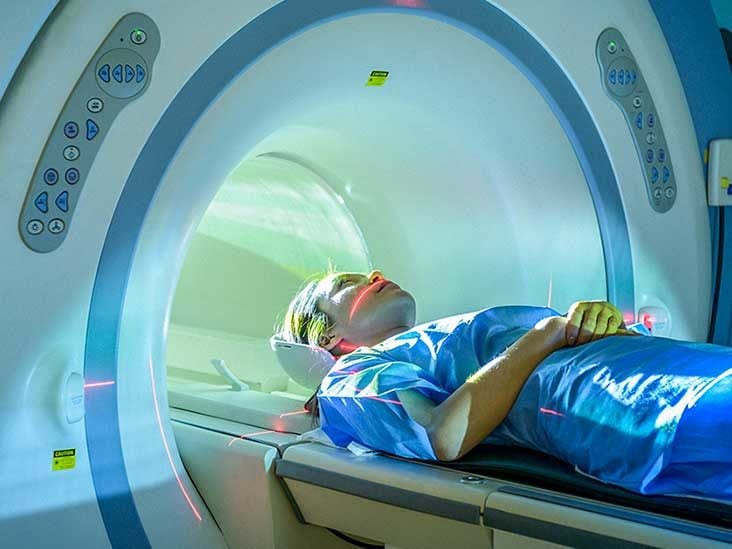
Brain PET Scan: Purpose, Procedure, and Risks
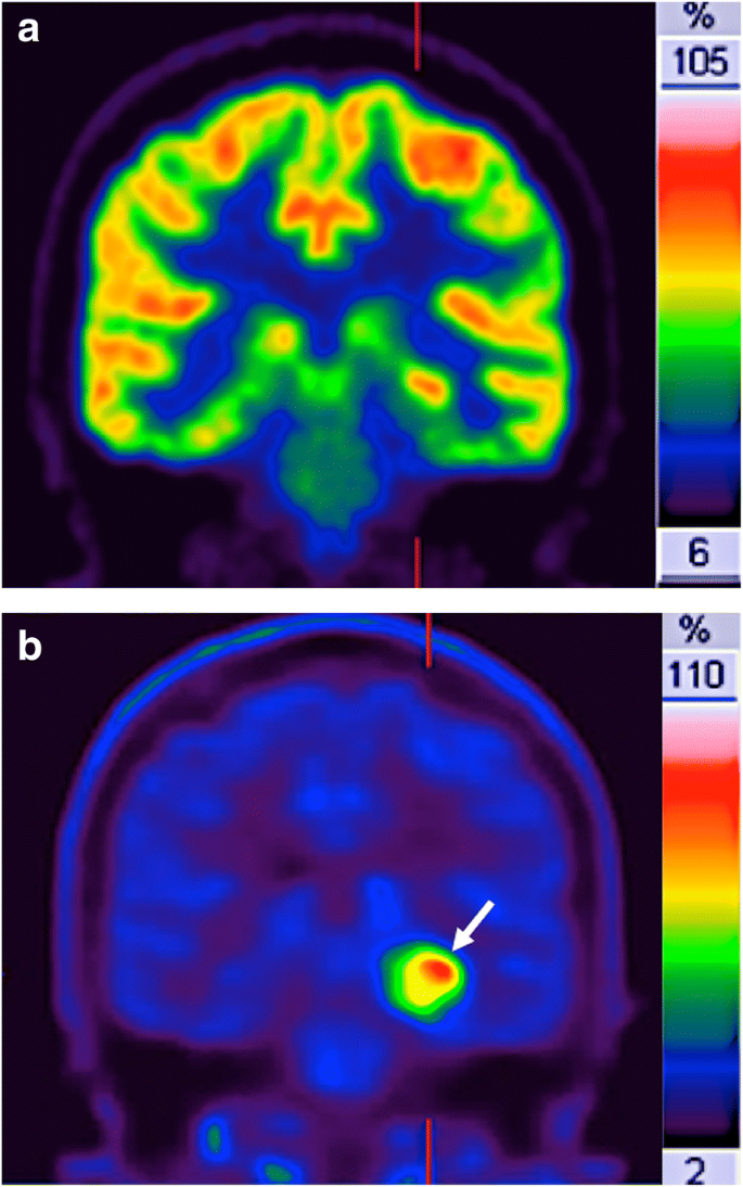
How clinical imaging can assess cancer biology | Insights into Imaging | Full Text
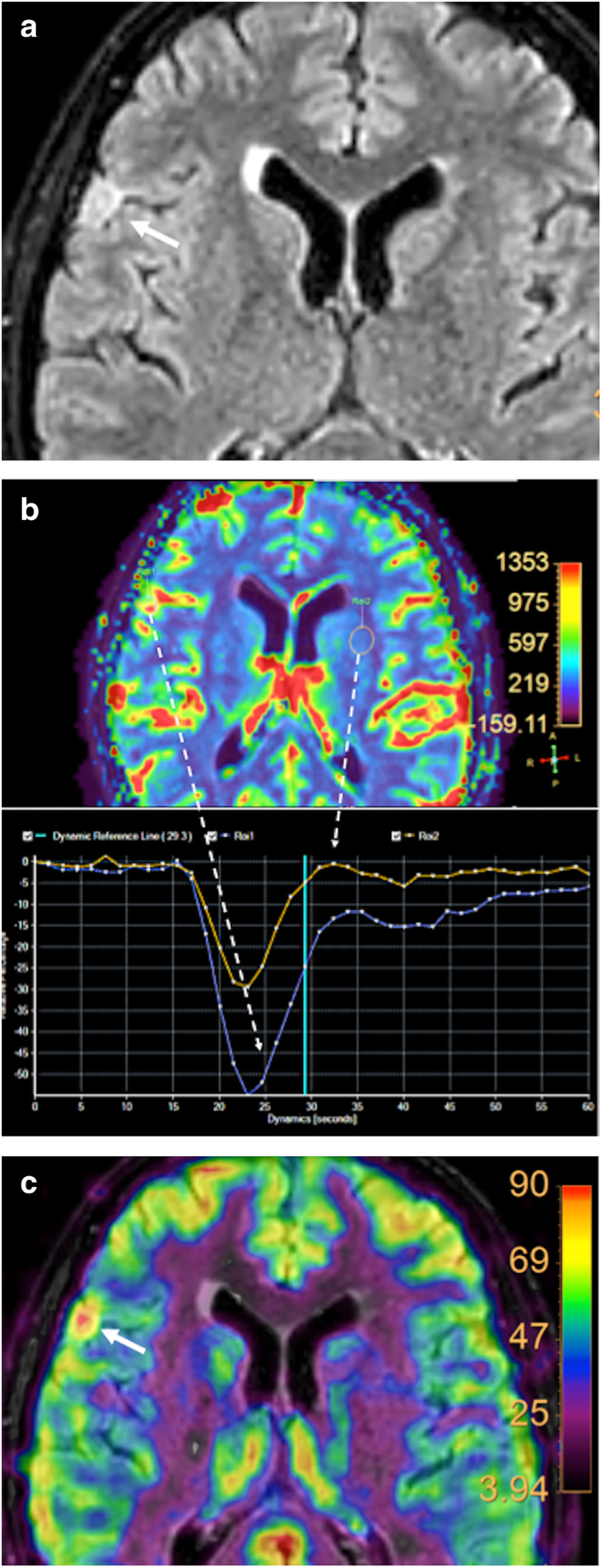
How clinical imaging can assess cancer biology | Insights into Imaging | Full Text
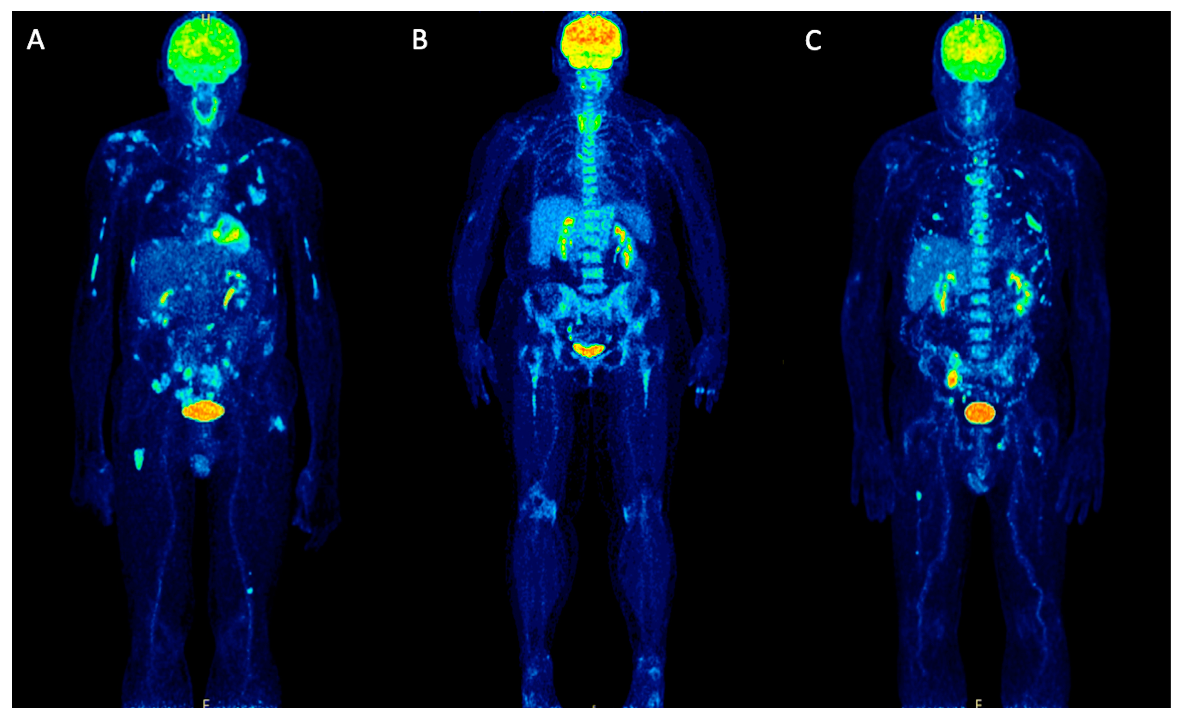
Molecules | Free Full-Text | Positron Emission Tomography (PET) Radiopharmaceuticals in Multiple Myeloma | HTML
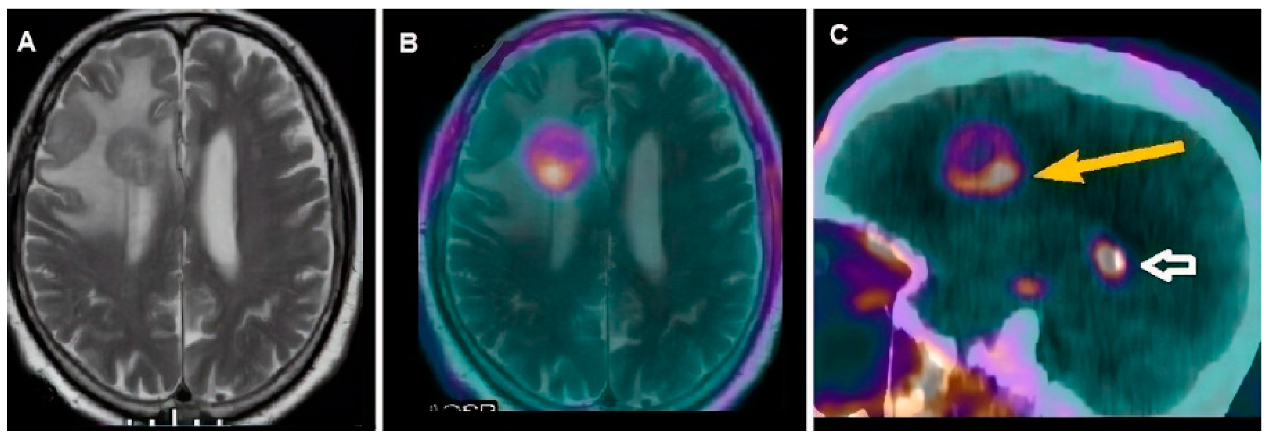
Cancers | Free Full-Text | Molecular Imaging in Pediatric Brain Tumors | HTML
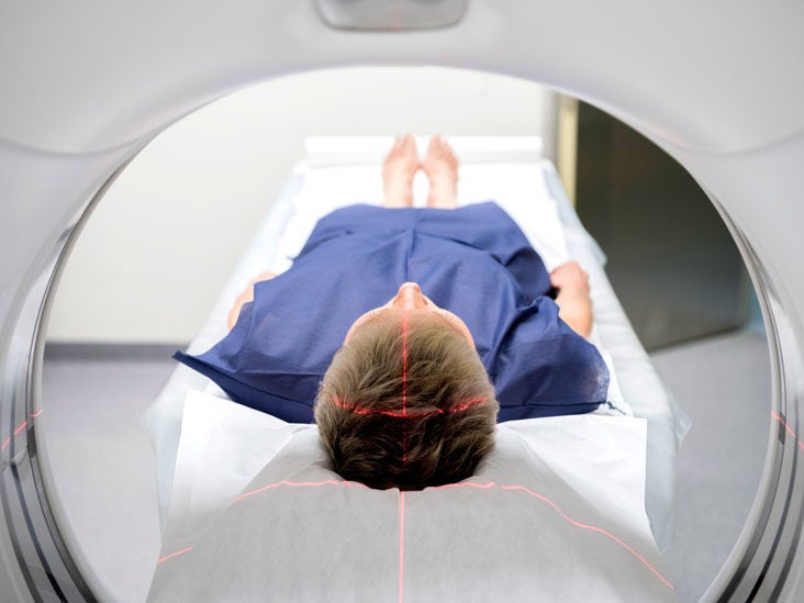
Brain PET Scan: Purpose, Procedure, and Risks
PET scan (positive emission tomograpy) information | myVMC

Brain Positron Emission Tomography - an overview | ScienceDirect Topics

Cancers | Free Full-Text | Molecular Imaging in Pediatric Brain Tumors | HTML
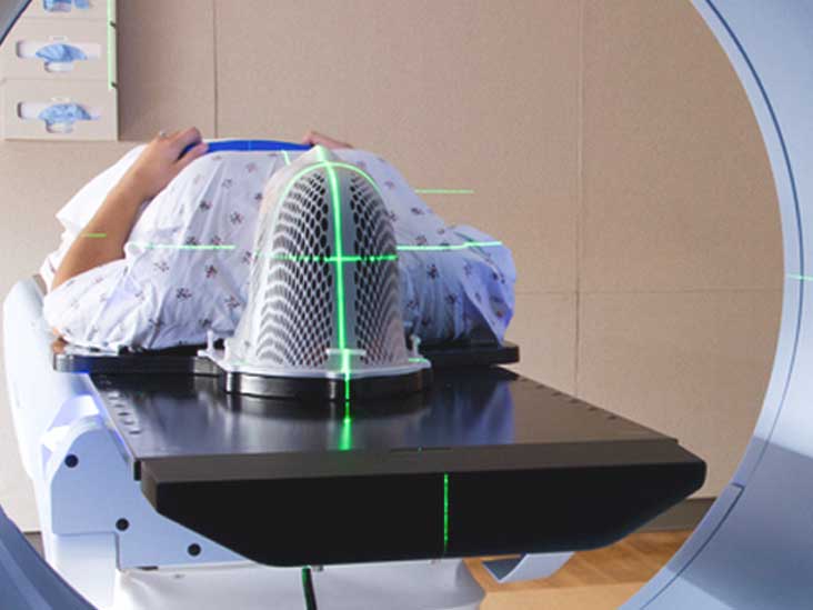
Brain PET Scan: Purpose, Procedure, and Risks

PDF) PET/MRI versus PET/CT in oncology: a prospective single-center study of 330 examinations focusing on implications for patient management and cost considerations

Differentiating Radiation Necrosis from Brain Tumor Using Hyperpolarized Carbon-13 MR Metabolic Imaging | SpringerLink
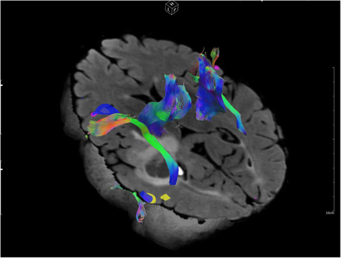
What scans we will read: imaging instrumentation trends in clinical oncology | Cancer Imaging | Full Text

Non-caloric sweetener provides magnetic resonance imaging contrast for cancer detection | Journal of Translational Medicine | Full Text
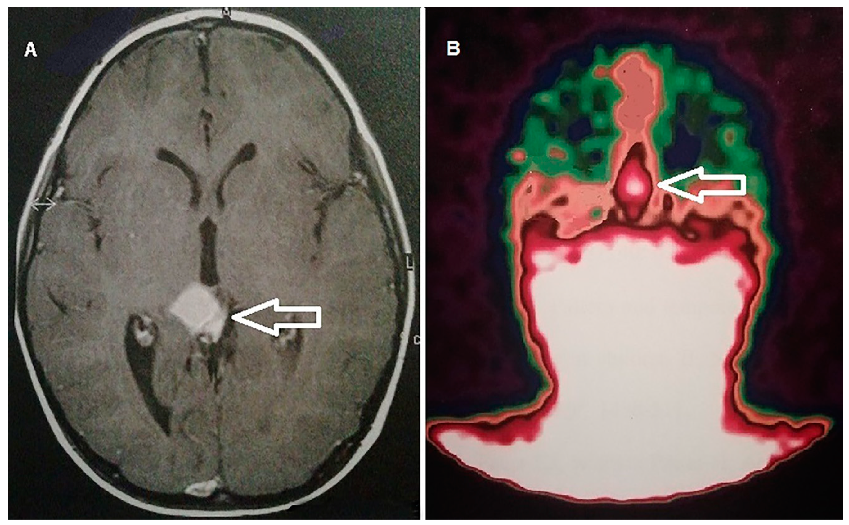
Cancers | Free Full-Text | Molecular Imaging in Pediatric Brain Tumors | HTML
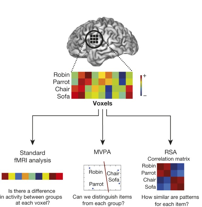
Progress and challenges in probing the human brain | Nature
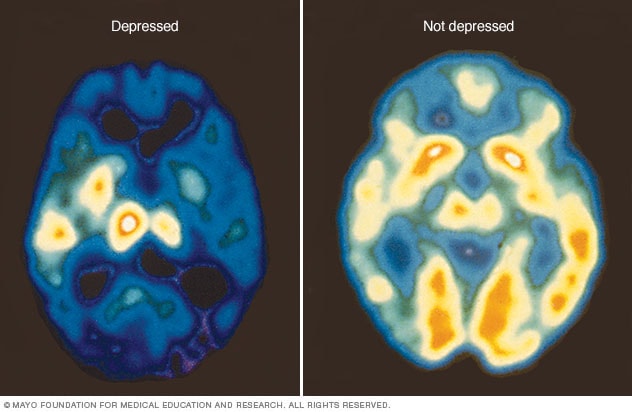
PET scan of the brain for depression - Mayo Clinic
MEG Frequency Analysis Depicts the Impaired Neurophysiological Condition of Ischemic Brain
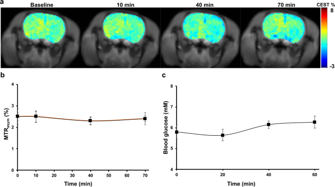
Sugar alcohol provides imaging contrast in cancer detection | Scientific Reports
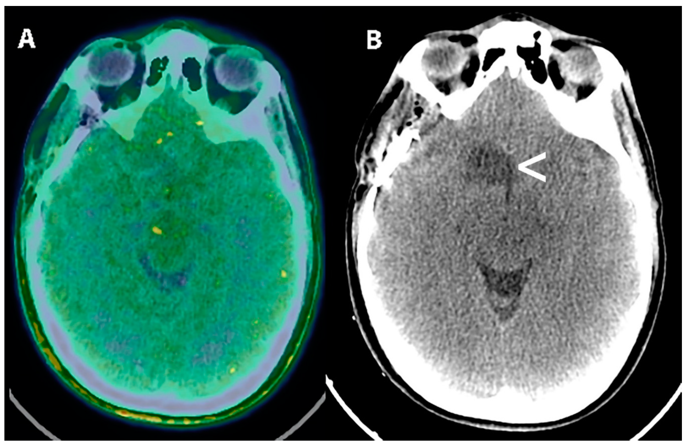
Cancers | Free Full-Text | Molecular Imaging in Pediatric Brain Tumors | HTML

PET-CT Principles and Applications in Lung Cancer Management | IntechOpen

Scanning the Brain

Brain Positron Emission Tomography - an overview | ScienceDirect Topics

Imaging of Pelvic Malignancies with In-Line FDG PET–CT: Case Examples and Common Pitfalls of FDG PET | RadioGraphics
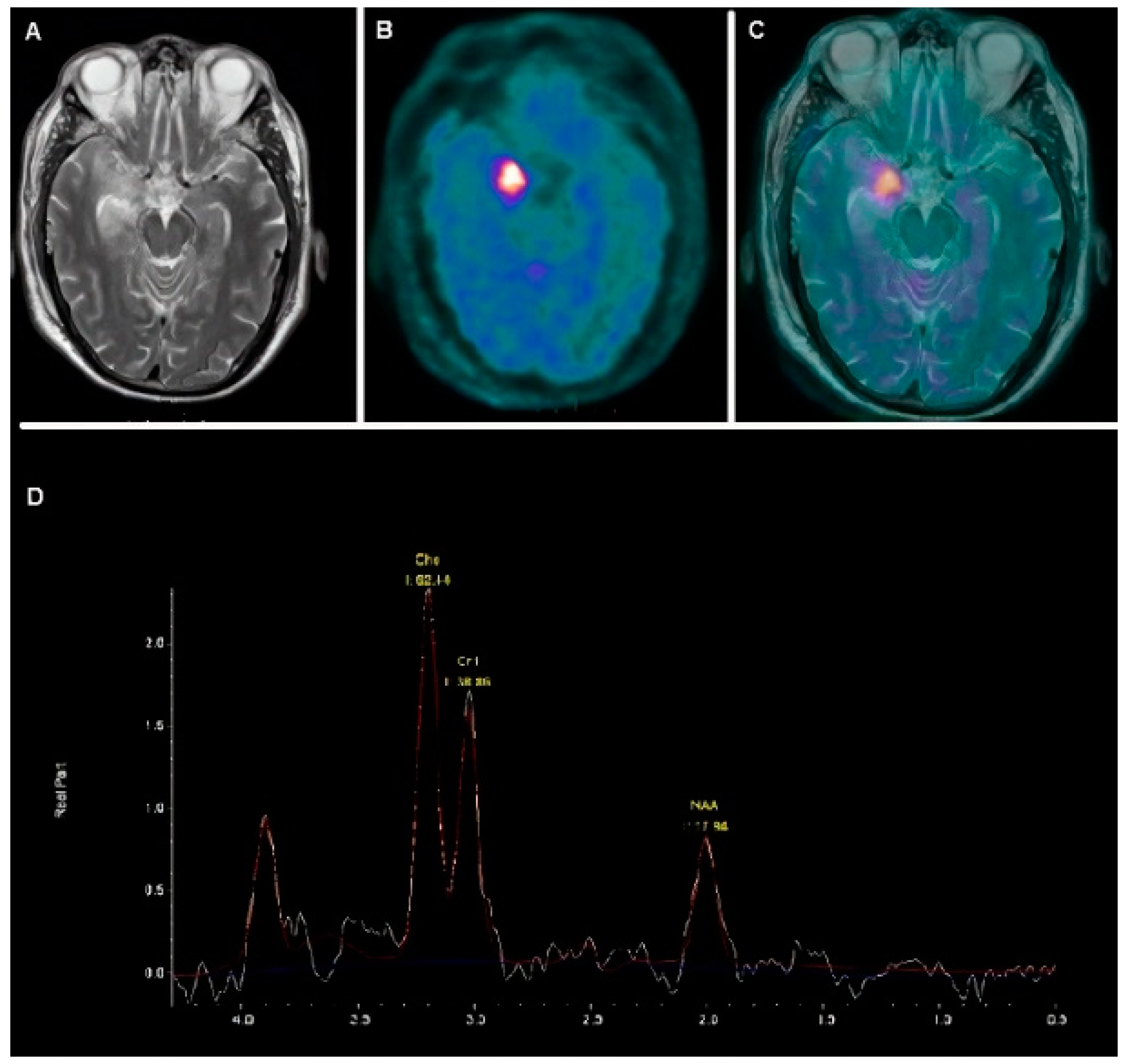
Cancers | Free Full-Text | Molecular Imaging in Pediatric Brain Tumors | HTML

Imaging of Pelvic Malignancies with In-Line FDG PET–CT: Case Examples and Common Pitfalls of FDG PET | RadioGraphics
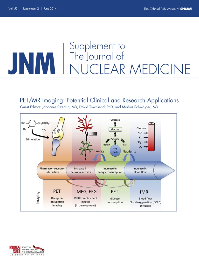
Potential Pediatric Applications of PET/MR | Journal of Nuclear Medicine
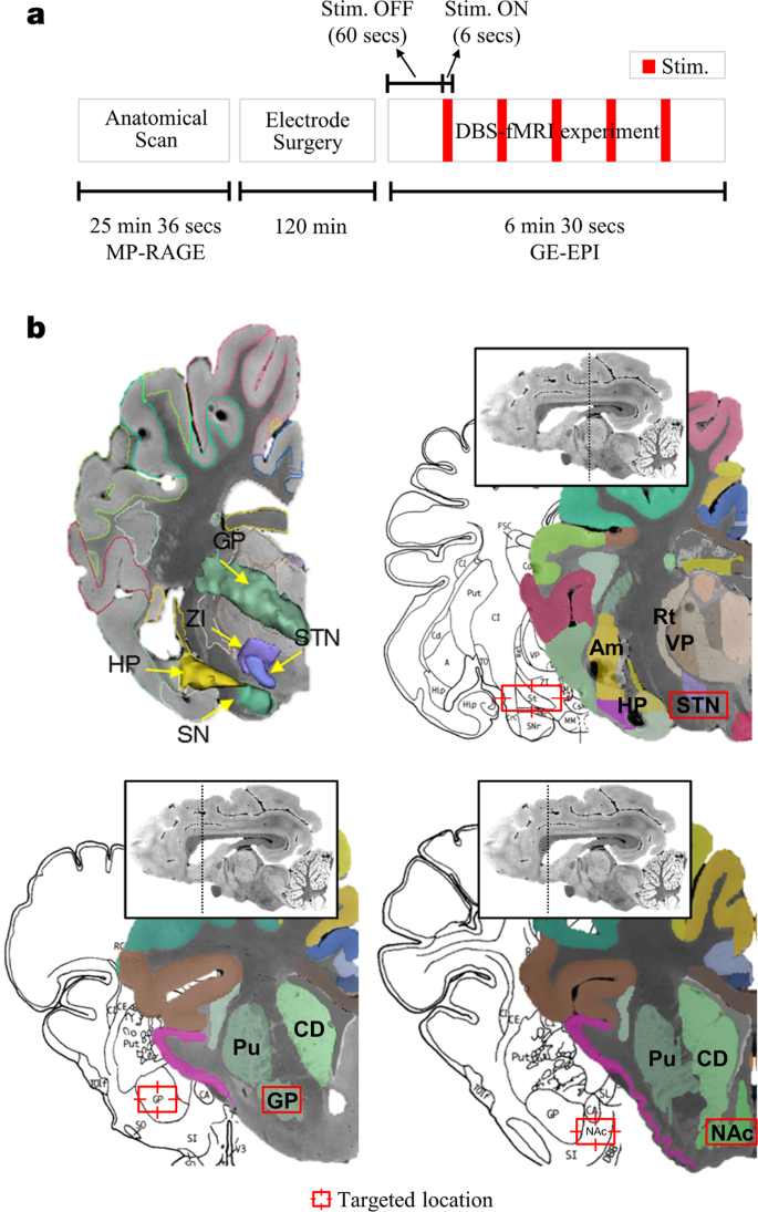
Multivariate pattern classification on BOLD activation pattern induced by deep brain stimulation in motor, associative, and limbic brain networks | Scientific Reports
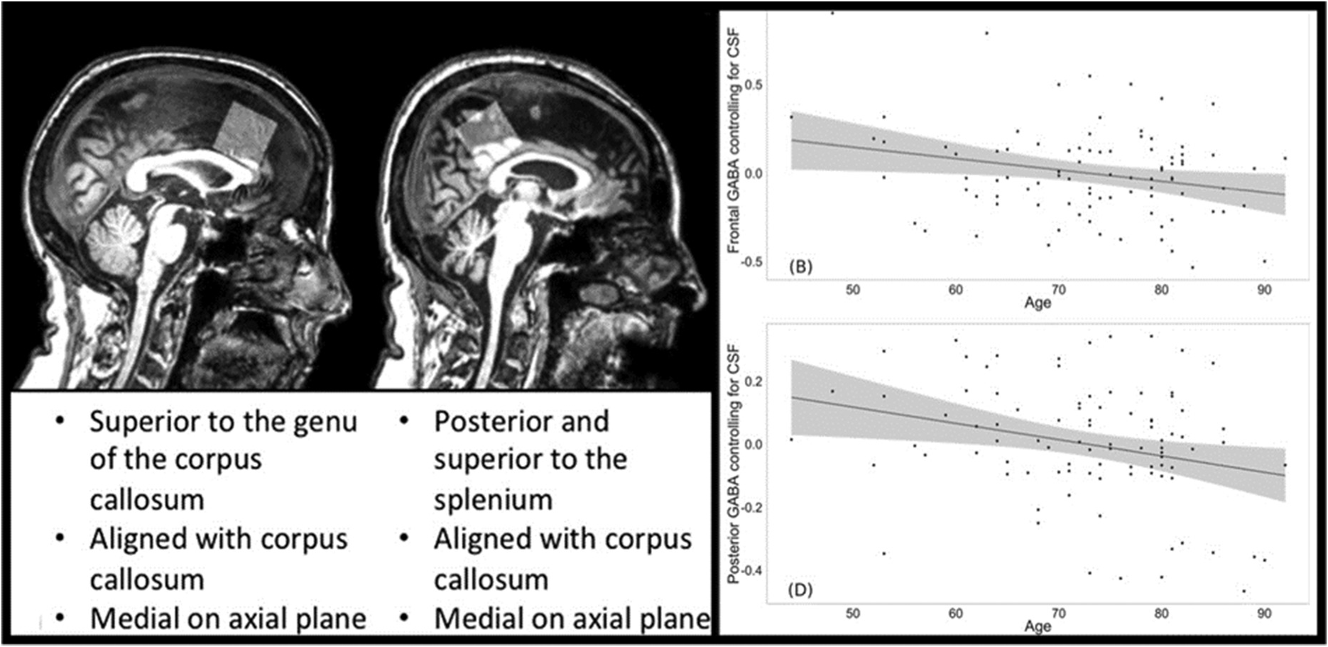
Neuroimaging of the Aging Brain (Chapter 4) - Cognitive Changes and the Aging Brain

Clinical utility of arterial spin labeling imaging in disorders of the nervous system in: Neurosurgical Focus Volume 47 Issue 6 (2019)

Brain PET Scan: Purpose, Procedure, and Risks

Projection Space Implementation of Deep Learning–Guided Low-Dose Brain PET Imaging Improves Performance over Implementation in Image Space | Journal of Nuclear Medicine
Usefulness of FDG PET/CT derived parameters in prediction of histopathological finding during the surgery in patients with pancreatic adenocarcinoma
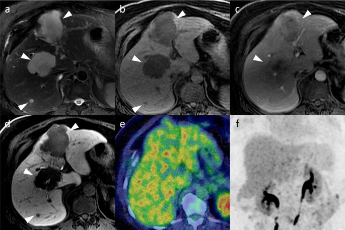
FDG-PET/CT imaging findings of hepatic tumors and tumor-like lesions based on molecular background | SpringerLink
![Quantification of brain oxygen extraction and metabolism with [15O]-gas PET: A technical review in the era of PET/MRI - ScienceDirect Quantification of brain oxygen extraction and metabolism with [15O]-gas PET: A technical review in the era of PET/MRI - ScienceDirect](https://ars.els-cdn.com/content/image/1-s2.0-S1053811920306224-gr6.jpg)
Quantification of brain oxygen extraction and metabolism with [15O]-gas PET: A technical review in the era of PET/MRI - ScienceDirect

Foundations of Human Consciousness: Imaging the Twilight Zone | Journal of Neuroscience

Study design. Overview on the study design for monitoring the influence... | Download Scientific Diagram
Posting Komentar untuk "a positron emission tomography pet scan depicts brain activity by detecting:"